
Most patients go to a neurosurgeon expecting an expert in their condition. However, while a doctor may be a brilliant surgeon, their knowledge of complex Ehlers-Danlos Syndromes (EDS) comorbidities can rank dangerously low.

Traditional Chiari decompression has an estimated failure rate approaching 50%. When a surgeon claims a “100% success rate,” be highly cautious. They are usually measuring mechanical completion (the bone was removed, the patch was sewn in) rather than how the patient actually feels.

Furthermore, data from our community of over 10,000 members reveals a deeply deceptive tactic used to protect these perfect statistics: these surgeons systematically cherry-pick their cases, dismissing complex patients before surgery—and entirely abandoning them if they develop a post-decompression problem afterward.

When a hypermobile patient experiences a post-op slump, worsening fluid pressure, or progressive instability, these surgeons routinely gaslight them, claim the operation was a structural success, and tell them to find a new neurosurgeon when post-decompression problems appear. By erasing injured patients from their clinics, they erase them from their failure data.

We deserve better than a tidy post-op scan followed by medical abandonment. Use the interview script below to find out if your surgeon understands a crowded skull versus a prolapsing brain.


  • Defining the Anatomy:
    How do you differentiate between a true congenital Chiari malformation and an Acquired Chiari Malformation driven by connective tissue laxity? Since the standard diagnostic criteria for Chiari is based entirely on the size of the tissue prolapse (herniated tonsils) rather than the physical size of the skull cavity, an MRI report for a congenital crowded skull and an Acquired prolapsing brain will look completely identical. How are you ruling out the latter before operating?
  • Volumetric Data:
    Are you basing my diagnosis solely on the millimeter drop of my cerebellar tonsils? Did my radiologist measure the physical volume of my posterior fossa to prove my skull cavity is actually too small?
  • The Prolapsing Risk:
    If my posterior fossa volume is normal, but my ligaments are fragile, how will performing a decompression prevent my hindbrain from simply prolapsing further into the newly enlarged opening?
  • The Chiari 1.5 Distinction:
    Looking at my sagittal scans, is any part of my medulla oblongata (brainstem) herniated or elongated below the foramen magnum? If yes, how does a basic decompression address this altered brainstem position?

  • The Core Measurements:
    What are my exact measurements for the Clivo-Axial Angle (CXA), Grabb-Oakes, and Harris measurements? [1]
  • Dynamic Imaging:
    Since instability is a motion failure, will you order Upright Dynamic (Flexion/Extension) MRI or Digital Motion X-Ray (DMX) to calculate how many millimeters my skull translates when moving? [2]
  • The Subaxial Foundation (C3–C7):
    Have you evaluated my mid-to-lower neck for Subaxial Instability (SAI) and premature Degenerative Disc Disease (DDD)? If my lower neck column is unstable or collapsing, how will my upper neck withstand the stress of a skull-base decompression? [4]
  • The Full-Spinal & Brain Scan Blindspot:
    If you are ordering a cervical scan but refusing a brain scan (or vice versa), how can we accurately map my neuro-anatomy? Since the central nervous system is one continuous column, how can we safely plan an irreversible surgery without a complete visualization of my brain down to my lumbar spine?
  • The Syrinx Origin:
    If I have a Syringomyelia (syrinx), how are we ruling out localized spinal cord compression from C3–C7 instability as the true driver of the cavity, rather than a skull-base blockage alone? If you refuse to scan my thoracic and lumbar spine, how can we be sure there isn’t a secondary syrinx or a tethering point fueling this fluid buildup? [4]

  • Addressing the Occult:
    Are you experienced in diagnosing Occult Tethered Cord Syndrome (OTCS)? If my conus medullaris terminates at a “normal” level on a standard supine MRI, how are we ruling out a tight, stretched cord that is anchoring my nervous system? [3]
  • Advanced High-Resolution Imaging:
    Will you order a CISS (Constructive Interference in Steady State) MRI sequence of my lumbar spine? Since standard thick-slice MRIs miss micro-structural tension, will we use a CISS sequence to check for a thickened filum terminale or nerve roots pinned against the spinal canal walls? [3]
  • Prone Imaging Dynamics:
    Will you order a Prone Lumbar MRI (lying on your stomach) alongside the CISS sequence to see if my spinal cord is pathologically anchored and fails to float forward normally? [4]
  • Medullary Stretching & Dysautonomia:
    Are my profound autonomic symptoms (POTS, blood pressure crashes) coming from direct brainstem compression, or is a tethered cord stretching my medulla oblongata from the bottom up? [3, 4]
  • Surgical Order of Operations:
    If I have an occult tethered cord pulling my brain down, why should we decompress my skull before releasing the bottom tension? Won’t the rubber-band pull simply drag my brainstem deeper into the empty surgical site?

  • The S.E.E.P.S. Scan Review:
    Do you see any of the classic signs of spontaneous intracranial hypotension (SIH) on my brain MRI? [5]
    • S — Subdural fluid collections?
    • E — Enhancement of the pachymeninges (brain lining)?
    • E — Engorgement of dural venous structures?
    • P — Pituitary hyperemia (enlargement)?
    • S — Sagging of the brain/brainstem?
  • The Invisible Fistula:
    Are you aware that CSF-Venous Fistulas (CVFs) act like an open drain, siphoning my brain downward, yet leave zero fluid pools on standard spinal MRIs? [5]
  • Advanced Imaging Verification:
    If I have a clear orthostatic (positional) headache but a “normal” MRI, will you refer me to a specialized CSF leak center for a Digital Subtraction Myelogram (DSM) or Ultrafast CT Myelogram to find the hidden fistula or leak before operating on my head? [6]

  • High-Pressure Structural Signs:
    Does my brain MRI show an Empty Sella (or partially empty sella), flattened lateral ventricles, or distension of the optic nerve sheaths? [7]
  • Venous Stenosis Assessment:
    Since dural sinus collapse is highly prevalent in the EDS population, will you order a Magnetic Resonance Venogram (MRV) to evaluate my internal brain drainage? [7]
  • The Outside-In “Eagle” Compression:
    Because I have a hypermobile neck, will you order a 3D-CT Reconstruction of my skull base to rule out Eagle Syndrome? Are my elongated or calcified styloid processes mechanically crushing my internal jugular veins when I turn my head? [7, 8]
  • The Post-Op Slump Risk:
    If my brain is being pushed down by high pressure from a blocked vein or Eagle Syndrome, how will a decompression fix my drainage? Won’t the high pressure simply shove my brain stem directly into the newly created space? [8]
  • Cranial Valve Check:
    If my high pressure has caused a Cranial CSF Leak (fluid leaking from nose or ears), why should we patch the leak before clearing the venous blockage that caused the pressure to blow out in the first place?

If a neurosurgeon answers defensively, dismisses your measurements, or attempts to schedule an immediate decompression while ignoring met diagnostic criteria in Track 1, 2, 3, or 4: STOP.

An untreated comorbidity is the primary driver of a failed surgery. Walk away, protect your anatomy, and find a surgeon who treats the human being, not just the herniation.

You are informed, empowered, and absolutely Worth the Fight (WTF!)


Oropharyngeal dysphagia = when your upper esophageal sphincter, pharynx, larynx, and/or tongue fail to coordinate properly when trying to swallow.

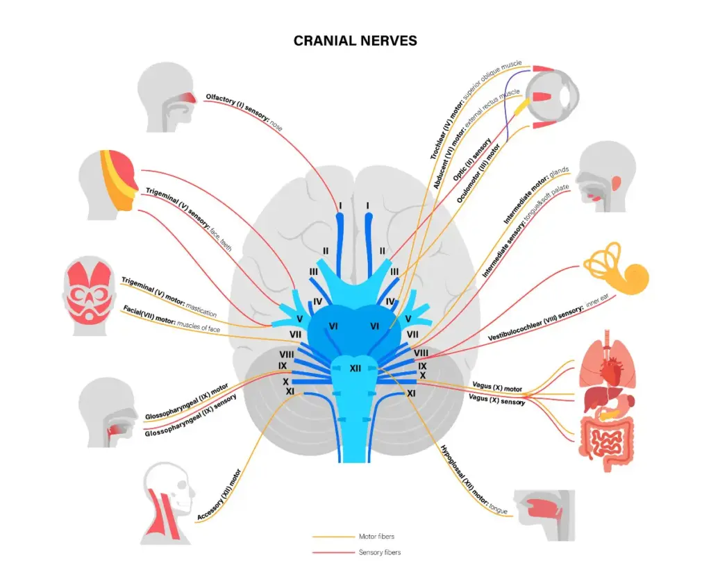
Can occur when one or more of the following nerves is damaged or compressed: facial nerve (CN VII), glossopharyngeal nerve (CN IX), vagus nerve (CN X), and hypoglossal nerve (CN XII).

Now that you know it’s a thing… ask your doctor for a swallow test to confirm it.

Neurosurgeons on this list are ones that have focused much of their practice on Chiari Malformation and Comorbid Conditions. Not everybody will agree with who is on this list and that’s okay. While we do encourage members to share THEIR OWN EXPERIENCES WITH THEIR DOCTORS, we DO NOT ALLOW DOCTOR BASHING in our groups! The doctors that have concerns (some of which are serious/major concerns), amongst patients in our group/site, we have marked with a (!) after their name. It might be worth asking in the group for those that have had negative experiences to share with you about what happened through private message.


ADULTS:

Dr. Petra Klinge – Rhode Island, USA
https://brownneurosurgery.com/our-team/petra-klinge-md-phd/
593 Eddy Street, APC 6, Providence RI, 02903; Ph. 1(401)793-9139 or 1(401)793 9166
Specializes in Tethered Cord and Occult Tethered Cord. *No remote consultations.

Dr. Fraser Henderson – Maryland, USA
http://metropolitanneurosurgery.org/practice/henderson.html
Spine Team Maryland 8116 Good Luck Road, Suite 205 Lanham, MD 20706; Ph. 1(301)654-9390
Specializes in Problems of the Craniocervical Junction (CCI/AAI/BI). Does craniocervical fusions. Insurance might be an issue. *No remote consultations.

Dr. Paolo A. Bolognese (!) – New York, USA
https://nspc.com/physician/paolo-bolognese/
Chiari Neurosurgical Center at NSPC 1991 Marcus Avenue, Suite 108 Lake Success, NY 11042
Ph. 1(516)321-2586; email pbolognese@chiarinsc.com
Specializes in Complex Chiari & Problems of the Craniocervical Junction (CCI/AAI/BI). Does craniocervical fusions. *Known to do video consults for $300. To initiate consultation for 2nd opinion, contact
Jeffrey.Wood@snch.org.

Dr. Gerald Grant – North Carolina, USA
https://med.stanford.edu/profiles/gerald-grant
Duke Children’s Health Center Neurosurgery Clinic, 2301 Erwin Rd, Durham, NC 27710-4699
Ph. 1(919)668-8557 or 1(919)684-5013
Chief of NS. Specializes in Pediatric NS > Chiari & Fusions.

Dr. Anthony L. Capocelli (!) – Arkansas, USA
http://www.orthoarkansas.com/capocelli.php
Ortho Arkansas – Orthopedics & Sports Medicine 10301 Kanis Rd. Little Rock, AR 72205
Ph. 1(501) 604-6900 (800) 264-5633
Neurosurgeon specializing in Chiari. Does craniocervical fusions.

Dr. Dan S. Heffez (!) – Wisconsin, USA
https://www.heffezchiari.com/
Private Practice Milwaukee, WI
Ph. 1(414)955-7188
Neurosurgeon claiming to specialize in Chiari.

Dr. Holly Gilmer (!) – Michigan, USA
https://www2.mhsi.us/doctors/holly-gilmer
Michigan Head and Spine Institute 29275 Northwestern Hwy., Suite 100, Southfield, MI, 48034
Ph. 1(248)784-3667
Neurosurgeon claiming to specialize in Chiari.


PEDIATRICS:

Dr. Douglas L. Brockmeyer – Utah, USA
https://healthcare.utah.edu/fad/mddetail.php?physicianID=u0034828
The University of Utah Health 50 North Medical Drive, Salt Lake City, UT 84132
Ph: 1(801)581-2121 douglas.brockmeyer@hsc.utah.edu
Specializes in Pediatric NS > Chiari & Fusions.

Dr. Richard C. Anderson – New Jersey, USA
https://www.valleyhealth.com/doctors/richard-c-anderson-md
Valley Hospital Health System 1200 East Ridgewood Ave Suite 200, Ridgewood, NJ 07450 Ph: 1(201)327-8600, 1(212)305-0219, Joanna (nurse) 1(973)758-3616 Rca24@cumc.columbia.eds
Specializes in Pediatric NS > Chiari (not sure about fusions).

Dr. Jeffrey Greenfield (!) – New York, USA
https://weillcornell.org/jpgreenfield
Weill Cornell Brain & Spine Center 1305 York Avenue, 9th Floor New York, NY 10021
Ph. 1(212) 746-2363
Specializes in Pediatric NS (but does take adults) > Chiari, some comorbids, does not do fusions, but contracts them out to another surgeon. Known to do remote online for $300 and phone consults for $500.


OTHER SPECIALTIES:

Dr. Wouter Schievink: LEAK SPECIALIST (NS) – California, USA
https://www.cedars-sinai.edu/Patients/Programs-and-Services/Neurosurgery/Centers-and-Programs/Cerebrospinal-Fluid-Leak/
Cedars-Sinai Advanced Health Sciences Pavilion, 127 South San Vicente Blvd. Suite A6600 Los Angeles, CA 90048
Ph. 1(310)423-3277 Wouter.schievink@cshs.org
Neurosurgeon who specializes in leaks. Known to review MRIs for free in patients with low pressure symptoms.

Dr. Ian Carroll: SPINAL LEAKS (EBP, Anesthesia, PM) – California, USA
Stanford 430 Broadway St., Pavilion C, RM 462; MC 6343, Redwood City, California 94063
Ph. 1(650)721-7286 (office) ic38@stanford.edu
Not a neurosurgeon, but his daughter had an Acquired Chiari secondary to a spinal leak, and therefore is VERY PASSIONATE about trying to find leaks in patients dx’d with Chiari if they have low pressure symptoms.

Dr. Kenneth C. Liu: VASCULAR NS – Michigan, USA
https://www.bronsonhealth.com/doctors/kenneth-liu/
Bronson Neuroscience Center – Kalamazoo 601 John St., Suite M-124 Kalamazoo, MI 49007
Ph. 1(269) 341-7500 kenneth.c.liu.md@gmail.com
Specializes in Neuroendovascular Surgery, Venous Hypertension, Intracranial Hypertension


RETIRED/NO LONGER TAKING PATIENTS

Dr. John Oró
https://auroramed.com/service/ccc
Colorado Chiari Institute – The Medical Center of Aurora 1501 S. Potomac Street Aurora, CO 80012
Ph. 1(303)695-2663 ChiariCare@HealthONEcares.com
RETIRED AS SURGEON, but oversees Colorado Chiari Institute

Dr. Faheem A. Sandhu
https://www.medstarhealth.org/doctor/dr-faheem-akram-sandhu-md/
MedStar Georgetown University Hospital – Neurosurgery 10401 Hospital Drive, Suite 101 Clinton, MD 20735
Ph. 1(301) 856-2323
NO LONGER ACCEPTING EDS PATIENTS

Hospitals and imaging centers in the United States are required to give you a copy of your imaging if you request it.

Many hospitals and imaging centers will give a copy of your MRI on disk or flash drive immediately after your appointment, but they do this as a courtesy and not as a requirement. (Keep in mind that you will still have to wait for a copy of your radiology report and might need to ask for a copy of that separately.)

Those that refuse to give you a copy immediately following your appointment must have procedures in place on how to obtain them. It will generally involve you, or your caretaker with medical power of attorney, contacting the facility’s records department and filling out a specific request form. Many states allow them to charge a minimal charge (usually no more than a few dollars), but most facilities will give at least one free copy. Some can have it ready in a matter of hours after making the request, while others can take up to two to three weeks. Some are willing to mail it to your home and others will require you to pick it up at their Patient Records Dept. (Different countries have different requirements, but most modern countries have procedures in place to request a copy.)

In the United States, records of imaging are legally allowed to be destroyed after a period of time (usually 7 years or less, depending on the state). For this reason, we recommend that you get a copy of all available MRIs and keep a copy for yourself and because disks are so easily destructible, we encourage you to back all of the contents up onto a hard drive and then store the disk away for safe-keeping. Never send anyone the original disk. Make copies as needed and send the copies.

If you decide to post your MRIs for Nonprofessional Opinions (NPOs) at WTF, please make sure that your post/images adhere to the following guidelines. Requests that do not meet our guidelines will be removed by an admin.

PLEASE MAINTAIN 100% PRIVACY TOWARDS THOSE TRYING TO HELP YOU OR SOMEONE ELSE. 
We have very strict privacy rules in this group. All images and comments given should be kept in the group or used privately by the owner of the image. Some members that give NPOs are okay with their input being shared, but it is the responsibility of the owner of the image to get permission from the person giving the NPOs before sharing them and all names should be edited out of all screenshots. Violating this rule will most likely result in an IMMEDIATELY removed/blocked from the group/site.

IMAGES SHARED MUST BE YOURS OR BELONG TO SOMEONE IN YOUR IMMEDIATE FAMILY.
Members are not allowed to share the images of their friends outside of this group. Violating this rule will be considered a breach of our privacy rules and will result in the member being IMMEDIATELY removed/blocked from the group/site.

WE DO NOT ALLOW PHOTOGRAPHS OF MRIs.
Photographs of images often contain glares, slants, and other problems that can compromise the image. We want you to get the best information possible here and we don’t want people going on a wild goose chase because the images were compromised in the first place. That will only make your doctors further disregard what you bring up to them.

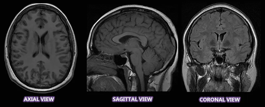
IN GENERAL, WE PRIMARILY PREFER SAGITTAL VIEWS:

  • Sagittal views are slices taken from one side to the other, viewing it from the left perspective. While sagittal is our preference, other views can be helpful as well:
  • Coronal views for instance are slices taken from the front to the back (so the left is really the right and vice-versa) and if you find the right slice, it can show if one cerebellar tonsil is lower than the other (it is also better for showing scoliosis).
  • Axial views are slices from the top to the bottom, so they can be instrumental in finding leaks (especially when contrast was used), but since a leak can be cranial or spinal and can happen anywhere along the way, you have to go through them closely one slice at a time.

ALL IMAGES MUST BE PROPER MIDSLICES:
When we talk about midslices, we’re NOT talking about the middle slice on the disk (although it is often close to that), we’re talking about the image showing the middle section of your head/neck/spine.

What you need to look for:

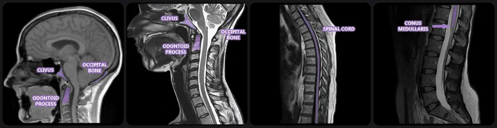
  • A midslice of the brain (example) should clearly show an unobstructed view of three bones: the clivus, the occipital bone, and the odontoid process.
  • A midslice of the cervical spine (example) should show the same three bones, but they will be slightly higher on the image.
  • A midslice of the thoracic spine (example) should show the vertebrae and full spinal cord. When scoliosis is present, you will see the spinal cord clearly at one point and then almost disappear (and sometimes you will see it come back into clear view – depending on the location of the curves of the scoliosis).
  • The spinal cord does not go as low as the vertebrae in the spinal column. The point in which it ends is called the conus medullaris and from there what continues down into the lumbar region is called the filum terminale. A midslice of the lumbar spine (example) should show the point where the spinal cord ends (conus medullaris) and the filum terminale begins.

*We do make one exception to images being midslice and that is if there is the presence of a mass (cyst/tumor) or lesion (any of which can be anywhere in the brain and may not be visible in the midslice). If that is the only image given, that is the only thing that we will be commenting on (we will not pretend that it is a midslice and comment on it as though it was).

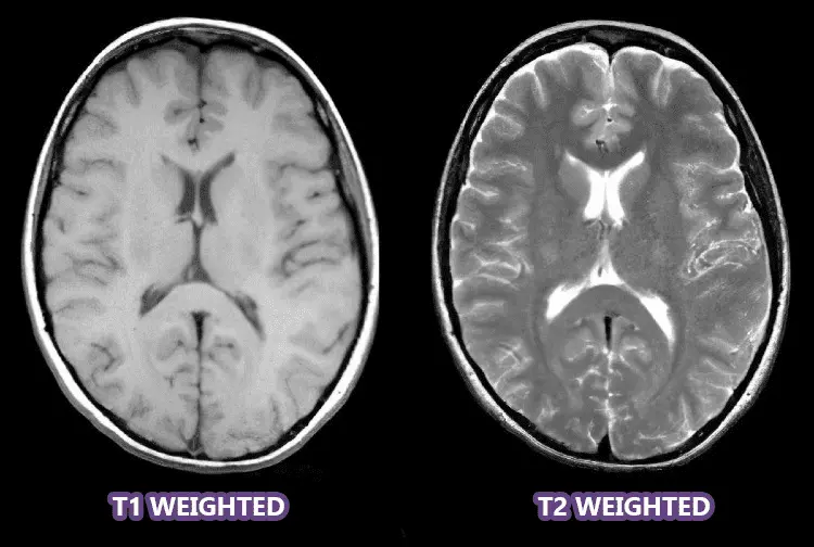
T2 WEIGHTED IS PREFERRED, BUT T1 IS PERMITTED AS WELL:

  • On a T1 weighted image, the cerebrospinal fluid (CSF) is dark gray or black.
  • On a T2 weighted image, the cerebrospinal fluid (CSF) is light gray or white. Having the CSF show up as white creates a better contrast that makes it easier to see certain things (such as intervertebral disc issues).

Image Size:
Small images make it hard for us to see what we’re looking for and even harder for us to try and mark what we see. Therefore, all images submitted should be cropped into a minimum of 500-800px. To accomplish this, please open the images full screen on your PC and either save them to size or use your snipping tool to snip them while they are large. Please DO NOT simply enlarge the picture as it reduces resolution that can compromise what can be seen.

NPO Markup with the Admin Think Tank:
The Nonprofessional Opinion (NPO) Request Form is required for all markup requests with the Admin Think Tank (no exceptions). Once you’ve submitted the form, you will be added to the waiting list (which is in our Facebook group), and an admin will contact as your name gets close to the top of the list.


HOMEPAGE: <https://chiaribridges.org/> (go to article)

ABOUT CHIARI (TAB)
  • Understanding Your Head and Neck Pain: <https://chiaribridges.org/understanding-your-head-and-neck-pain/> (go to article)

  • Overview: Chiari Malformation [Revised]: <https://chiaribridges.org/chiari-malformation/> (go to article)
  • What’s In A Name? An Expansive Review of the Name and Definition of Chiari Malformation: <https://chiaribridges.org/whats-in-a-name-chiari-malformation/> (go to article)

  • One Painful Fight – Get Ready To Rumble!!!: <https://chiaribridges.org/painful-fight-get-ready-rumble/> (go to article)

  • The Diagnosis – Round One: <https://chiaribridges.org/diagnosis-round-one/> (go to article)

  • The Important Questions to Ask Your Neurosurgeon [Revised]: <https://chiaribridges.org/important-questions-for-your-neurosurgery-appointment/> (go to article)

  • Overview: Chiari Comorbidities & Etiological/Pathological Cofactors [Revised]: <https://chiaribridges.org/overview-chiari-comorbidities-etiological-pathological-cofactors/> (go to article)

  • Overview: Chiari Treatment Options & Potential Pitfalls: <https://chiaribridges.org/chiari-treatment-options-potential-pitfalls/> (go to article)

  • Overview: Complications Associated With A Chiari Decompression: <https://chiaribridges.org/complications-associated-with-chiari-decompression/> (go to article)

  • A Bruised Mind – Chiari & Depression: <https://chiaribridges.org/bruised-mind-depression/> (go to article)

  • Finding Hope In The Seemingly Hopeless Chiari Fight!: <https://chiaribridges.org/finding-hope-seemingly-hopeless-chiari-fight/> (go to article)
COMORBIDITIES (TAB)
  • Overview: Chiari Comorbidities & Etiological/Pathological Cofactors [Revised]: <https://chiaribridges.org/overview-chiari-comorbidities-etiological-pathological-cofactors/> (go to article)

  • The Chiari Malformation Ehlers-Danlos Connection: <https://chiaribridges.org/chiari-malformation-ehlers-danlos-connection-2/> (go to article)

  • Overview: Craniocervical Instability and Related Disorders: <https://chiaribridges.org/craniocervical-instability-related-disorders/> (go to article)

  • Brain Under Pressure – A Guide to Understanding Intracranial Hypertension:<https://chiaribridges.org/brain-under-pressure-a-guide-to-understanding-intracranial-hypertension/> (go to article)

  • Overview: Cerebrospinal Fluid Leaks: <https://chiaribridges.org/cerebrospinal-fluid-leaks/> (go to article)

  • Spine Pulled Tight – A Guide to Understanding Tethered Cord Syndrome: <https://chiaribridges.org/spine-pulled-tight-a-guide-to-understanding-tethered-cord-syndrome/> (go to article)
WARRIOR STORIES (TAB)
  • Listing of stories: <https://chiaribridges.org/warriors/> (go to article)

  • The Michelle Cole Story – A Chiari Warrior’s Journey: <https://chiaribridges.org/the-michelle-cole-story-a-chiari-warriors-journey/> (go to article)
MISCELLANEOUS
  • Donations: <https://chiaribridges.org/donate/> (go to article)

  • Volunteer: <https://chiaribridges.org/join-the-cb-volunteer-team/> (go to article)

  • Share Your Story: <https://chiaribridges.org/submit-your-story/> (go to article)

  • Submission Agreement: <https://chiaribridges.org/submission-agreement/> (go to article)

  • The Chiarian’s Glossary: <https://chiaribridges.org/glossary/> (go to article)
Chiaribridges

FREE
VIEW