

Make the Most of Your Initial Appointment
Most patients go to a neurosurgeon expecting an expert in their condition. However, while a doctor may be a brilliant surgeon, their knowledge of complex Ehlers-Danlos Syndromes (EDS) comorbidities can rank dangerously low.
Traditional Chiari decompression has an estimated failure rate approaching 50%. When a surgeon claims a “100% success rate,” be highly cautious. They are usually measuring mechanical completion (the bone was removed, the patch was sewn in) rather than how the patient actually feels.
Furthermore, data from our community of over 10,000 members reveals a deeply deceptive tactic used to protect these perfect statistics: these surgeons systematically cherry-pick their cases, dismissing complex patients before surgery—and entirely abandoning them if they develop a post-decompression problem afterward.
When a hypermobile patient experiences a post-op slump, worsening fluid pressure, or progressive instability, these surgeons routinely gaslight them, claim the operation was a structural success, and tell them to find a new neurosurgeon when post-decompression problems appear. By erasing injured patients from their clinics, they erase them from their failure data.
We deserve better than a tidy post-op scan followed by medical abandonment. Use the interview script below to find out if your surgeon understands a crowded skull versus a prolapsing brain.
Part 1: The Foundation (Crowded Skull vs. Prolapsing Brain)
- Defining the Anatomy:
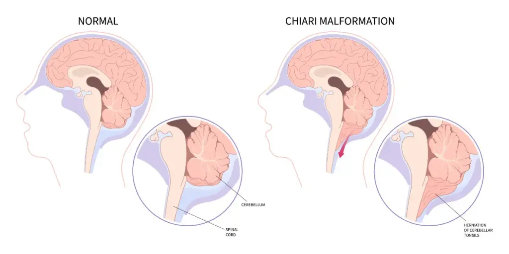
How do you differentiate between a true congenital Chiari malformation and an acquired Chiari Malformation driven by connective tissue laxity? Since the standard diagnostic criteria for Chiari are based entirely on the size of the tissue prolapse (herniated tonsils) rather than the physical size of the skull cavity, an MRI report for a congenital crowded skull and an acquired prolapsing brain will look completely identical. How are you ruling out the latter before operating?
- Volumetric Data:
Are you basing my diagnosis solely on the millimeter drop of my cerebellar tonsils? Did my radiologist measure the physical volume of my posterior fossa to prove my skull cavity is actually too small?
- The Prolapsing Risk:
If my posterior fossa volume is normal but my ligaments are fragile, how will decompression prevent my hindbrain from prolapsing further into the newly enlarged opening?
- The Chiari 1.5 Distinction:
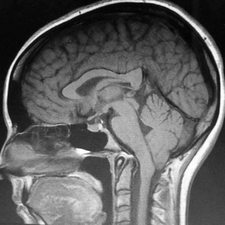
Looking at my sagittal scans, is any part of my medulla oblongata (brainstem) herniated or elongated below the foramen magnum? If yes, how does a basic decompression address this altered brainstem position?
Part 2: Track 1 — The Cervical Spine Instability Pathway
- The Core Measurements:
What are my exact measurements for the Clivo-Axial Angle (CXA), Grabb-Oakes, and Harris measurements? [1]
- Dynamic Imaging:
Since instability is a motion failure, will you order an Upright Dynamic (Flexion/Extension) MRI or digital motion X-ray (DMX) to calculate how many millimeters my skull translates when moving? [2]
- The Subaxial Foundation (C3–C7):
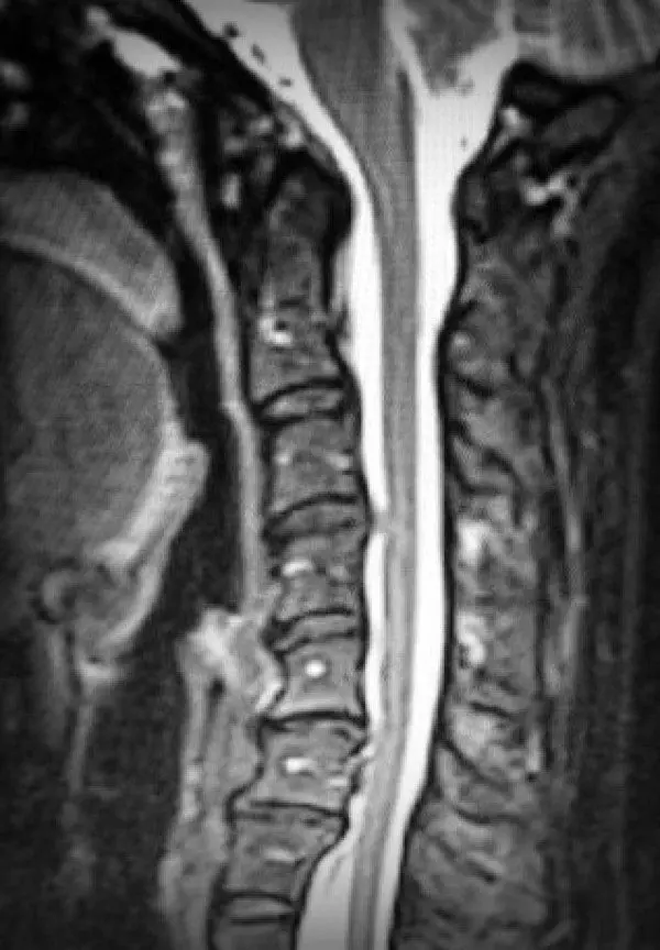
Have you evaluated my mid-to-lower neck for Subaxial Instability (SAI) and premature Degenerative Disc Disease (DDD)? If my lower neck column is unstable or collapsing, how will my upper neck withstand the stress of a skull-base decompression? [4]
- The Full-Spinal & Brain Scan Blindspot:
If you are ordering a cervical scan but refusing a brain scan (or vice versa), how can we accurately map my neuro-anatomy? Since the central nervous system is one continuous column, how can we safely plan an irreversible surgery without a complete visualization of my brain down to my lumbar spine?
- The Syrinx Origin:
If I have a Syringomyelia (syrinx), how are we ruling out localized spinal cord compression from C3–C7 instability as the true driver of the cavity, rather than a skull-base blockage alone? If you refuse to scan my thoracic and lumbar spine, how can we be sure there isn’t a secondary syrinx or a tethering point fueling this fluid buildup? [4]
Part 3: Track 2 — The Traction “Pull” Track
- Addressing the Occult:
Are you experienced in diagnosing Occult Tethered Cord Syndrome (OTCS)? If my conus medullaris terminates at a “normal” level on a standard supine MRI, how are we ruling out a tight, stretched cord that is anchoring my nervous system? [3]
- Advanced High-Resolution Imaging:
Will you order a CISS (Constructive Interference in Steady State) MRI sequence of my lumbar spine? Since standard thick-slice MRIs miss microstructural tension, will we use a CISS sequence to check for a thickened filum terminale or nerve roots pinned against the spinal canal walls? [3]
- Prone Imaging Dynamics:
Will you order a Prone Lumbar MRI (lying on your stomach) alongside the CISS sequence to see if my spinal cord is pathologically anchored and fails to float forward normally? [4]
- Medullary Stretching & Dysautonomia:
Are my profound autonomic symptoms (POTS, blood pressure crashes) coming from direct brainstem compression, or is a tethered cord stretching my medulla oblongata from the bottom up? [3, 4]
- Surgical Order of Operations:
If I have an occult tethered cord pulling my brain down, why should we decompress my skull before releasing the bottom tension? Won’t the rubber-band pull simply drag my brainstem deeper into the empty surgical site?
Part 4: Track 3 — The Low-Pressure “Siphon” Track
- The S.E.E.P.S. Scan Review:
Do you see any of the classic signs of spontaneous intracranial hypotension (SIH) on my brain MRI? [5]- S — Subdural fluid collections?
- E — Enhancement of the pachymeninges (brain lining)?
- E — Engorgement of dural venous structures?
- P — Pituitary hyperemia (enlargement)?
- S — Sagging of the brain/brainstem?
- The Invisible Fistula:
Are you aware that CSF-Venous Fistulas (CVFs) act like an open drain, siphoning my brain downward, yet leave zero fluid pools on standard spinal MRIs? [5]
- Advanced Imaging Verification:
If I have a clear orthostatic (positional) headache but a “normal” MRI, will you refer me to a specialized CSF leak center for a Digital Subtraction Myelogram (DSM) or Ultrafast CT Myelogram to find the hidden fistula or leak before operating on my head? [6]
Part 5: Track 4 — The High-Pressure Push Track
- High-Pressure Structural Signs:
Does my brain MRI show an Empty Sella (or partially empty sella), flattened lateral ventricles, or distension of the optic nerve sheaths? [7]
- Venous Stenosis Assessment:
Since dural sinus collapse is highly prevalent in the EDS population, will you order a Magnetic Resonance Venogram (MRV) to evaluate my internal brain drainage? [7]
- The Outside-In “Eagle” Compression:
Because I have a hypermobile neck, will you order a 3D-CT Reconstruction of my skull base to rule out Eagle Syndrome? Are my elongated or calcified styloid processes mechanically crushing my internal jugular veins when I turn my head? [7, 8]
- The Post-Op Slump Risk:
If my brain is being pushed down by high pressure from a blocked vein or Eagle Syndrome, how will a decompression fix my drainage? Won’t the high pressure simply shove my brain stem directly into the newly created space? [8]
- Cranial Valve Check:
If my high pressure has caused a Cranial CSF Leak (fluid leaking from nose or ears), why should we patch the leak before clearing the venous blockage that caused the pressure to blow out in the first place?
THE MASTER ADVOCACY RULE
If a neurosurgeon answers defensively, dismisses your measurements, or attempts to schedule an immediate decompression while ignoring met diagnostic criteria in Track 1, 2, 3, or 4: STOP.

An untreated comorbidity is the primary driver of a failed surgery. Walk away, protect your anatomy, and find a surgeon who treats the human being, not just the herniation.
You are informed, empowered, and absolutely Worth the Fight (WTF!)