Chiari 1 Malformations, Explained

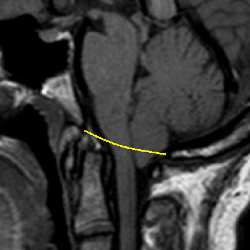
The most common type of Chiari Malformation, Type 1 is diagnosed when the cerebral tonsils descend below the foramen magnum, but the brainstem does not.
Some radiologists believe that a tonsillar herniation of less than 5mm is simply a tonsillar ectopia within normal limits, and only diagnose a Chiari Malformation when the descent is >5mm. However, the 5mm requirement is controversial, and many doctors have now moved away from diagnoses based solely on measurements, but rather on symptomology and a combination of other factors.[1] Some doctors will require a cine MRI to offer further evidence that the herniated cerebellar tonsils are blocking the flow of cerebrospinal fluid. This can sometimes be beneficial to convince an insurance company that has otherwise declined surgical coverage.
Many people with a Chiari Zero or Chiari 1 malformations can be asymptomatic for a lifetime: one large study found that approximately 30% of those with a CM measuring between 5-10mm were asymptomatic.[2] If symptoms develop, they often present in adolescence or early adulthood. Anecdotal evidence supports the proposition that once symptoms start, the symptoms often progress rapidly until the damage is stopped surgically.
Diagnosis Requirements: Symptomology; midsagittal MRI indicating at least one herniated tonsil (without the brainstem descending as well).[3]
Being that there are two bilateral cerebellar tonsils, it is important to note that 96% of patients have asymmetric tonsils, meaning one side is herniated more than the other (the right side tending to be lower than the left). This can lead to misdiagnoses when the doctor puts more weight in the midsagittal view than he ought. Instead, a coronal view can be used to compare the overall symmetry of the tonsils.[4]
Treatment Options: Any/all comorbidities should be explored and treated (if plausible) prior to surgery. If you are found to have a normal sized posterior fossa and no other pathological comorbidities that might be attributing to it, a decompression might be the logical next step. The risks of surgery should be weighed against the severity of symptoms and the impact that symptoms are having on the patient’s quality of life. It is often recommended to treat mild symptoms with medication, with surgical options typically reserved for cases in which symptoms cause more serious medical and quality of life problems. However, symptoms do tend to progress, and studies have shown a correlation between successful decompression surgery and the amount of time between the onset of any symptoms and surgical intervention[5]. See additional articles linked below.
Related Articles:
- Overview: Chiari Comorbidities & Etiological/Pathological Cofactors
- Understanding Your Head and Neck Pain
- The Important Questions to Ask Your Neurosurgeon
- Overview: Chiari Treatment Options & Potential Pitfalls
- Overview: Complications Associated With A Chiari Decompression
References:
1 Isik, N, et al. “A New Entity: Chiari Zero Malformation and Its Surgical Method.” Turkish Neurosurgery., U.S. National Library of Medicine, <www.ncbi.nlm.nih.gov/pubmed/21534216>.
2 Elster, A D, and M Y Chen. “Chiari I Malformations: Clinical and Radiologic Reappraisal.”Radiology., U.S. National Library of Medicine, May 1992, <www.ncbi.nlm.nih.gov/pubmed/1561334>.
3 Wilson, Eugene. “Chiari.” CEDSA Home, <www.cedsa.org/index.php/59-quick-reference/73-chiari.html>.
4 “The Chiari Malformations.” edited by R. Shane Tubbs et al., 2nd ed., 9 June 2020.
5 Hindawi. “Surgical Management of Patients with Chiari I Malformation.” International Journal of Pediatrics, Hindawi, 28 June 2012, <www.hindawi.com/journals/ijpedi/2012/640127>.
